Histiocytosis (LCH)
There are generally first signs of LCH and frequently become manifest as scaly,
erythmatous, seborrhea like brown to red papules, especially pronounced in inter-triginous
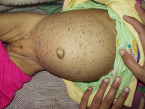
zones. Hepatosplenomegaly is the indication of presence of organ involvement
by LCH or it may indicate obstructive disease caused by enlarged nodes in the
porta hepatis. It may also reflect kupffer cell hypertrophy and hyperplasia
as an indicator of generalized activation of the cellular immune system. Cough,
tachypnea/dyspnea, cyanosis, pneumothorax, or pleural effusion are attributable
to the disease rather than to superimposed infection. Increasing number of cysts
form honeycomb lungs and in later stages large bullae. This child had fever
and was pale. The seborrhoeic dermatitis lesions were seen over scalp and abdomen.
He had cervical lymphadenopathy. Abdomen was soft on palpation with liver 8
cm below costal margin and spleen 4 cm below costal margin. Preliminary blood
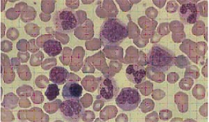
counts show hemoglobin 6.5 gm%, TLC 6300, Platelet count 68,000, DLC N49 L46
M02 My01 MM02.